Homepage Star of November 2015
Facial asymmetry can be caused by various factors. Among them the most common causative factors include the improper habitual such as constantly unilateral chewing or habitual torticollis, and heritage conditions such as muscular torticollis. It frequently involves both bone and soft tissue asymmetry of the face. It is very rare for facial asymmetry that only involves soft tissue or bone alone. There are different manifestation of facial asymmetry. However most facial asymmetry sufferers have facial midline deviation and tilted occlusion plan. For the correction of facial midline deviation the best choice of surgery is the bimaxillary osteotomy. For those whose facial midline is not deviated a differentiation ostectomy of the mandible bone and zygomatic bone can make the face more symmetric. As the bimaxillary osteotomy surgery is more complex than a simple reduction of the zygomatic and mandible bone some of the patients who suffer only minor midline deviation also tried the differentiated bone removal for the correction of asymmetric face with success.
The girl on this page had an asymmetric face with unknown etiology. Physical examination rolled out muscular torticollis. Her facial midline deviation was minor and her occlusion plan was only slightly tilted. For her the above mentioned bimaxillary surgery was too much. So she selected differentiated ostectomy of the mandible bone and zygomatic bone. The surgery focused on reducing the right zygomatic bone and left mandible bone plus the left masseter muscle. At the half year post-surgery follow up her facial asymmetry was greatly improved as shown in the following photos. The girl herself was satisfied with the result.

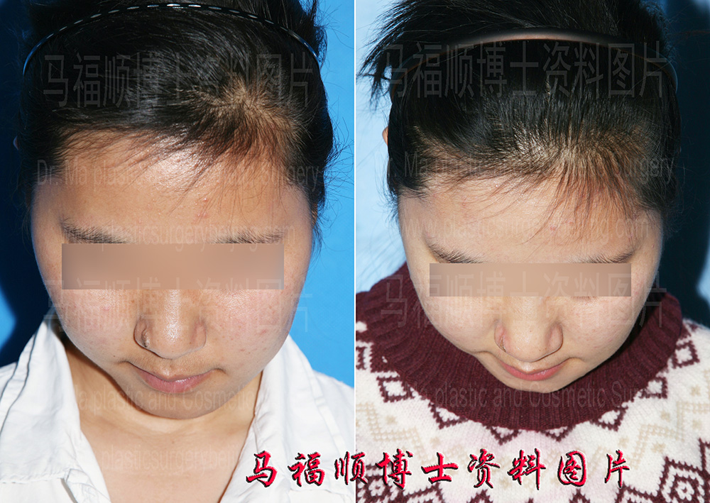
Facial asymmetry correction with differentiated ostectomy of the mandible bone and zygomatic bone., before and after surgery photos.

The bigger and saggy right side of the face was reduced and lifted after surgery.

In chin down position the two sides of this patient’s face look more symmetric in the post surgery photo on the right side.
The right side of this girl’s face is much bigger than the left side in the before surgery photos. Her facial asymmetry was not caused by muscle torticollis or habitual unilateral chewing. The etiology of her facial asymmetry was unknown. Under close examination the left side of her face was generally bigger. However her zygomatic bone and zygomatic arch was more prominent on the right side. The left corner of her mouth was lower than the right side because of the oversized left face. The degree of mouth corner dropping on the left side overrun the tilt degree of her teeth occlusion plan. As she only had a minor facial midline deviation her occlusion plan was slightly tilted.
Comparing to the before surgery photos the facial asymmetry of this girl was obviously corrected. The facial sizes of both sides were balanced and the mouth corners were leveled. The right zygoma looks no more prominent after zygomatic bone and zygomatic arch reduction surgeries. In the left profile photos the size of the mandible bone and masseter muscle is reduced in the post surgery photos. In order to achieve balanced reduction effect the right zygoma was reduced accordingly. As a result the reflect area of the zygoma in the post surgery photo is smaller. In chin down position both side of the zygoma was reduced, however the right side was changed more remarkably.
For a better result this girl may need to reduce the width of the front part of her left mandible bone also known as chin bone. In the frontal and lateral view of the after surgery photos the side of the chin bone seems becoming even bigger than the opposite side. That is because the rear part of the mandible bone and the masseter muscle on this side was reduced the spared frontal part appears relatively bigger. The chin reduction surgery is simpler and safer compared to her previous surgeries.
The after surgery photos was taken only half year post surgery. At that time the scar underneath the incision still exist. The scar swelling still had some influence to the surgery result. More time was needed to allow the scar to subside. Then her face would be even more symmetric.
